Ayatana, Sanskrit āyatana (“field” or “base”), in Buddhist philosophy, the field of cognition. Human physical existence consists of 12 ayatanas: the 6 cognitive faculties (5 sense organs and the mind) and the 6 corresponding categories of objects.
From the 19th of September to the 26th I took part in the Infinitesimal Ayatana Research Program. Situated in the beautiful Gatineau forest (in Chelsea), in an equally lovely 200 year old, refurbished farmhouse overlooking the river. Our days were spent in research facilities, laboratories and museums located at Carleton University, Ottawa U and the Canadian Museum of Nature. Our days were shock full of learning, exploration and knowledge/inspiration gathering- in mass! I mention the Buddhist definition of Ayatana because that is exactly what this research program was about; igniting all the senses!
Day 1: I arrived in Ottawa via Porter airlines, took the bus to Byward market. We were scheduled to meet at the National Museum of Canada for noon. It would turn out that only 4 of us could make this residency. The fantastic 4! I have to say that out of all the residencies I’ve experienced so far, this one was the best concerning diversity of artist’s use of mediums, but we were all like-minded, focussed and hit it off instantly. Alexis Williams is the director of the Ayatana programs. We were each given pocket microscopes for our adventures ahead, and to keep!

Day 2: After a quiet fog filled morning (I woke up at 4am!) listening to insects and birds call in the sunrise, we had a DIY breakfast before heading into Ottawa to Carleton University. First up was visiting the Carleton Nano Imaging Facility (NIF), home to two state-of-the-art Electron Microscopes, Tescan VegaII XMU SEM and FEI Tecnai G2 TEM.

The tungsten Tescan VegaII SEM images specimen surface profiles and topographies at variable pressures. It is operational at high vacuum for maximum resolution or low vacuum for direct imaging of fresh unprocessed biological specimens. It is equipped with Oxford X-ray detection systems (INCA EDS) for elemental analysis and quantitative mapping. A Cryo stage is also available for the low-temperature operation.
The FEI Tecnai G2 TEM reveals specimen information down to the atomic level where it reaches a stunning 1-million magnification. The FEI Tecnai G2 features a field emission source which makes the ultra-high resolution imaging possible – a spatial resolution as small as 0.144nm can be achieved. The Tecnai G2 is equipped with the latest Oxford X-ray detection systems – Aztec EDS which offers high-degree accuracy at a fast data acquisition speed.
Physicist Jianquan Wang aka JJ introduced us to this amazing technology. We ‘plated’ and scanned a moth antennae, a fly, a moth head, silk cocoon, dragonfly wing, blue fly, and yarrow. 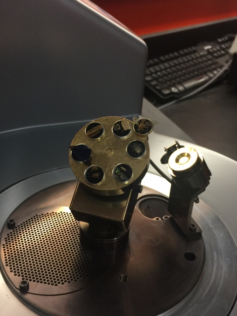


JJ was super enthusiastic and accommodating. He explained Back Scatter technology, the vacuum plating equipment, and demonstrated how to take samples (dry in this case) and prep them for plating. He is also very funny. 🙂 


JJ asked me if I sang opera 😉 We could have easily spent the entire day with JJ and his SEM, chatting sciart, the future, bio-art, etc., but we had another appointment scheduled with the Petrographic lab and Dr. Beth Halfkenny.

Looking a super thin slices of minerals is interesting. I never thought to look into Petrographic saws for cutting glass! SO, this visit was also incredibly inspirational. Also, looking at basalt (from Hawaii with Olivine inclusions) with a polarizing lens.. whoa.  I made several videos while moving the specimen in circles- I will post later. Dr. Halfkenny also shared this seriously cool microscope hack with us-
I plan to build one. On our way out.. again, we did not want to leave! She showed us some fossils she really loves.  This little guy and this most spectacular Crinoid. !!


This Jasper specimen (below) shows oxygen just starting to form on the Earth’s surface.


After this incredible day of science we headed to the LCBO then back to the house to decompress, write, discuss the day and share new exploding ideas!
Thanks so much for reading! Ayatana research residency pt2 coming soon…

Wonderful, wonderful, wonderful! Art On!